Do you know how your eye is build? |
| 09.10.2017 |
 Sight is one of the most important senses. After all, it is the source of approximately 80% of information about our immediate surrounding! As many as 10% of neurons in the brain take part in processing visual sensations. Thanks to the eye we are not only able to look but also to see – and understand the world around us.
The visual system consists of the eyeball (which receives optical input) visual tracts (which conduct the signal) and visual cortex (which is the main processor of visual input).
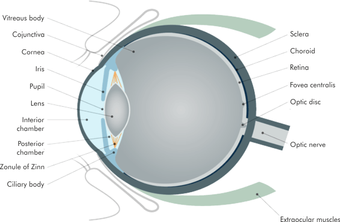
The eyeball is shaped like a sphere. It is approximately 24 millimetres and is located in the anterior part of the orbit. It is filled with aqueous humor – it is a pressured substance which helps eyeball maintain its spherical shape. The eyeball consists of three membranes – outer fibrous layer (cornea and sclera), middle vascular membrane (iris, uvea and ciliary body) and the inner layer retina). Cornea emerges from the sclera in its’ anterior part – they conform an elastic “scaffolding” of the eyeball. Behind the cornea is the iris, which gives our eyes their colour and is responsible (via constriction and dilation of the pupil) for regulating the amount of light entering the eyeball. If the light is too intense the pupil can change its’ width to 2 millimetres, while during the night it can become as wide as 8 millimetres. Retina is comprised of multiple layers of cells: pigment epithelium, neurons, glial cells and photoreceptor cells – rods and cones, the former are responsible for telling light from darkness while the latter enable us to perceive colour. The main function of the retina is perception of visual stimuli. Macula lutea is the area of the retina where most of the cones are concentrated, the rods are uniformly distributed throughout the whole are of retina. The macula ceca is located below – it is totally deprived of photoreceptor cells. Uvea is responsible for nutrition of the outer layers and the ciliary body holds the lens in place.

The lens is situated between the iris and the aqueous humor. It is second, after the retina, part of the eye responsible for refracting the incoming light. Thanks to the movement of the ligaments, which connect it to the iris and ciliary body it can modify its’ refracting power (this process is called accommodation – when we concentrate on far objects, the lens flattens, analogically it becomes more convex when we try to observe nearest surroundings).
The eyeball is moved by three muscle groups, which are responsible for rotating it in different axes. It is protected by the orbit, eyelids, eyelashes, conjunctiva and lacrimal apparatus. The orbit protects it from mechanical damage. In the posterior part of the orbit the opening of optic canal is situated (the optic nerve (which conducts visual impulses to the cortex). The eyelids and eyelashes protect the eye against excessive light as well as possible contamination and traumas. When we blink the eyelids distribute the tears, which are produced by the lacrimal apparatus. The tears are essential for the correct function of the eye – they dissolve and remove any harmful substances, also they have bacteriostatic and bactericidal properties.